What Scares You?

It’s well into the spooky season and Halloween just round the corner. With many of us indulging in a few scares and horror movies. I felt now was a thematic time to talk about fear. That primal emotion that causes goose-bumps, butterflies in our stomachs and sweat to drip down our necks. What is fear and how do we develop it?

At its base fear is a survival instinct. All things that cause us to be afraid, can in our minds lead us to death. That is the base of all fears we experience, the body’s natural need to survive and prevent anything that would do us harm, potentially leading to our demise.

The Amygdala

This region of the brain is responsible for controlling our response to the things that scare us. It receives vast amounts of sensory information, from our vision, hearing and sense of touch. The amygdala shares a close connection to another region of the brain known as the hippocampus (our memory centre), allowing the processing of past experiences or information to determine the threat level and therefore the strength of fear produced. It learns from experience, measuring the dangers of the world, and acts to trigger the fight or flight response. Allowing us to defend ourselves against imminent danger or to run like hell to get away from it. During the fight or flight response there is a huge release of adrenaline, causing increases in heart rate drawing blood to the muscles ready to act. Blood is also redirected away from less important processes to focus on the muscles. It is this diversion of blood that causes the feeling of butterflies, as your digestive system has a reduced supply.

Information from outside the body is communicated to the amygdala. This information is processed and compared with memories from the hippocampus. If a fear response is needed the amygdala triggers action from the hypothalamus (this region of the brain has many roles including aiding in the fight or flight response). This leads to adrenaline release from the adrenal gland.

Becoming Afraid

There are several innate fears we as humans possess. Scientists can categorise these into 6-8 distinct groups. We are born with two innate fears these are a fear of falling and loud noises. New-born babies surprisingly feel comfortable in the dark, as it reminds them of being in the womb. As we develop we gain other fears that all link to the pinnacle fear of death. These fears include:

  • Falling
  • Pain
  • Unknown – the abnormal, creepy faces or the dark.
  • Sudden Movement/Noise
  • Disease
  • Isolation – being alone or rejected. Linked to the fear of public speaking.
  • Suffocation

Many of the common fears people experience can be linked back to these base fears. Such as a fear of spiders can be connected to the sudden movements they exhibit or the potential chance of being bitten, hence pain aversion. Many fears are learned or inherited through one’s own experience or the stories from others. Fear can also be obtained vicariously, seeing another afraid can instil that same fear onto you. Understanding how fears develop was first shown in the real life horror story of the Little Albert Experiment.

A psychological study on fear was published in 1920. This was called the Little Albert Experiment. It set out to determine whether you could condition a phobia into an emotionally stable child. A nine-month old referred to as “Albert” was exposed to several different stimuli. They were introduced to a rat, a rabbit, a monkey and other furry things including cotton wool. As expected the baby showed no sign of fear to these things. The true experiment started at age 11 months. The child was placed upon a bed with a white rat and allowed to play with it. Though, each time Albert made contact with the rat the experimenters hit a metal bar with a hammer. This unpleasant loud noise being a natural fear to children was repeated each time Albert tried to play with the rat. Albert showed the signs of fear along with crying in response to this noise. Eventually Albert would recoil and retreat whenever the rat was brought close. The level of distress was then observed upon Albert seeing a rabbit and a dog, even a Santa Claus mask with a furry beard triggered fear in the child. Scientifically this was the first true demonstration of fear conditioning. However this experiment was morally wrong, the lead scientist John B Watson did not have the time to desensitise the child again and it is believed that the phobia continue post experiment. Modern fear studies are not conducted in this manner and work is performed to prevent any lasting harm.

The Woman with No Fear

The next story I shall cover is patient S.M, a woman from the USA whose identity is kept private for her own protection. S.M is incredibly unique, as at a young age she suffered with a condition called Urbach-Weithe disease which left her amygdala destroyed. She was later dubbed by the media the “woman with no fear.” S.M went through several studies proving that she was unable to experience being scared. Haunted attractions, scary movies, snakes, spiders were all met with curiosity or excitement rather than terror. She has been the victim of muggings at knife point to which she showed no behaviour response associated with panic or desperation. She has experienced an extraordinarily high number of life threating events, which is believed to be due to her inability to determine the danger of specific situations. Situations that could have been avoided. S.M is unable to truly recognise threatening behaviour from people and an inability to interpret aggressive facial expressions. Interestingly, there is something that scares even S.M. The brain is an incredible thing and doesn’t need to amygdala to know that something is terribly wrong causing panic to set in. S.M was given a ventilation mask and the amount of CO2 was increased generating the sensation of suffocation. Her reaction was immediate and extreme, for the first time in her life S.M was afraid. Something very crucial was learnt from this experiment on S.M. Another role of the amygdala in suppressing fear. Her reaction was very extreme, when other participants with functioning amygdala were tested they showed fear, but deep down knew that this experiment wasn’t going to kill them. They believed they were safe, S.M was unable to make this connection in her brain.

Getting Over Fear

Many of these irrational fears we as humans possess can be dealt with through therapy. Two major techniques have seen success in treating phobias. The first and most gentle method is through systematic desensitisation. Take for example the fear of spiders. Early on in therapy you may be asked simply to read a book about spiders, then move on to looking at pictures of them. You are fully aware that these images and words can’t harm you. This helps to unravel the connections you have made in your mind between spiders and death that your amygdala has set up. The next step would be to be in a room with a spider in a tank. The spider can’t get you it’s just there. Finally you would likely be asked to handle a spider. Over time this trains your brain that spiders don’t control your fear, and you can overcome it. The second therapy method is through flooding. This usually involves putting the fear front and centre. Day one placing a spider straight in your hands for example. An extreme method, but apparently quite effective.

Obviously medical therapeutics are also an option. Anti-anxiety drugs are effective for reducing the panic associated with fears. Drugs including diazepam or alprazolam (probably better known as Valium and Xanax respectively). However only tend to be prescribed in cases where the fears are irregular and unavoidable. Such as a fear of the dentist, flying or surgical procedures. Beta-blockers can also be used. These act to prevent the action of adrenaline, therefore avoiding the blood flow changes, heart rate increases and sweating.

Joy in Fear

Fear is a fascinating part of our brain. It aims to prevent us from death. However the world is so messy and memories impact how we perceive normally innocuous things. There are many of us that get enjoyment from being scared. Particularly when sharing it with others. Interestingly the state we enter during fear is not dissimilar to that of excitement. The major difference between these two states is the context through which we are experiencing them. Fear is triggered by danger and excitement through fun. There are many things that we deem fun that trigger fear and it is the knowledge that we are safe that turns that fear into joy. Fear can work as a natural high. The falling feeling and speed of rollercoasters can make the amygdala think we are in danger, but we know better so that rush of adrenaline is put to better use. This is the origin of the adrenaline junky. The seeking of scary situations for pleasure. So however you spend your Halloween, being scared isn’t all that bad.

Reading Sources

  • The Biology of Fear, Ralph Adolphs, 2014.
  • Retrieving fear memories, as time goes by…, Do Monte et al., 2017.
  • Mechanisms of Fear Learning and Extinction: Synaptic Plasticity – Fear Memory Connection. Luchkina & Bolshakov, 2020.
  • Fear and panic in humans with bilateral amygdala damage, Feinstein et al. 2013.
  • Impaired recognition of emotion in facial expressions following bilateral damage to the human amygdala. Adolphs et al. 2016.
Your Friendly Tenants

Just as we live on planet earth and make it our home, along with millions of other plants and animals. Some organisms have chosen to make our bodies their home. I am sure many will have seen or heard the term “good bacteria.” Most often used in health adverts, pushing us to buy drinks to improve or promote our gut microbiome. They are not wrong these bacteria that have set up residence in our intestines are in fact highly beneficial to us. They play a major role in digestion, helping us to break down food mater that we ourselves don’t have the ability to do. This provides us with normally inaccessible nutrients along with supplying themselves with food they need. These bacteria also as a by product of their own digestion provide chemicals that are beneficial to our health. For example some bacteria will produce butyrate, this works as an energy source for our intestinal cells and can in turn help prevent colon cancer.

Our microbiome is held in a delicate balance, thousands of different species all co-existing to benefit the whole. So when these bacteria are lost or one group out numbers another problems can begin. This can arise when people are on antibiotics for long periods of time. Leading to a knock on effect to the numbers of gut bacteria. To correct this imbalance some medical experts recommend the use of a rather unpleasant sounding technique. This would be the faecal transplant (you did read that right). The faeces of a healthy donor can be used to acquire bacterial colonies that are then reintroduced into the patient who suffered the loss. Thankfully we do not use the method of the past and drink the solution.

Gut Bacteria and Cancer

Why talk about gut bacteria now? We have known for a while that our residential bacterial are symbiotic (providing benefit to themselves and us). Recent work has shown how these bacteria are actually able to help us fight against cancerous tumours. The bacteria are able to augment our own immune system, helping it to identify and attack cancers such as melanoma, colorectal and bladder.

Coronavirus stress causing stomach troubles? Here are 6 tips to comfort  your gut at home - Times of India

One of the more recent techniques in treating cancer is through immunotherapy. This is a technique that aims to amplify your own immune system to attack the cancer. A group lead by Dr Kathy McCoy at the University of Calgary have been investigating the role that gut bacteria can play in assisting the host’s immune system in fighting cancer. We have know for some time that these bacteria can help fight cancer but the exact method was yet to be discovered. We now know through Dr. McCoy’s work that the bacteria “enhance the ability of the T-Cells” to attack and eventually destroy cancerous cells.

T-cells are white blood cells, the major players of the immune system. There are a large variety of T-cell from helper T-cells (which play a supportive role in the function of the immune system) to killer T-cells (whose name I feel is clear enough). One of the functions of killer T-cells is to recognise and attack cancerous tumour cells.

Dr. McCoy used immunotherapy in mice and then identified bacteria that had associated themselves with the colorectal cancer cells. Specifically Dr. McCoy used a technique called immune checkpoint blockade. This effectively removes the safety precautions the immune system has in place to not attack its own cells. As cancer cells are your own mutated cells, the immune system can sometimes ignore them, allowing them to grow and become tumours. This method helps the immune system detect the cancer cells more easily. The group were then able to combine the immune blockade with cultures of the associated bacteria leading to observed improvements in the therapy. The tumours shrank and in some cases the T-cells were able to destroy all the cancerous cells present. The group identified several bacteria that were linked to increasing the effectiveness of immunotherapy. These bacteria were producers of the chemical inosine. This had the effect of activating anti-tumour T-cells, which as their name suggests target tumour cells for death.

The group continued their work and showed the similar results with bladder cancer and melanoma. Next the research needs to be demonstrated in human tissues. Dr McCoy said “We are in the early stage of fully understanding how we can use this new knowledge to improve efficacy and safety of anti-cancer therapy and improve cancer patient survival and well-being.” This could be the beginnings of a huge step for cancer treatment. By utilising our own bacterial residents we could work together to save their home from cancer.

Reading Sources

  • Microbiome-derived inosine modulates response to checkpoint inhibitor immunotherapy. McCoy et al., Science, 2020.
  • The gut microbiome. Herb Brody, Nature, 2020.
  • Role of the gut microbiota in nutrition and health. Ana Valdes, Jens Walter, Eran Segal, Time Spector. The bmj, 2018.
  • Symbiotic Human Gut Bacteria with Variable Metabolic Priorities for Host Mucosal Glycans. Martens et al. 2015.
  • National Cancer Institute.

Parkinson’s Disease: Repairing the Brain

I have previously written about the developments in transplantation surgery. How we will eventually be able to replace tissues with self grown stem cells. Well that day has come, recent work has shown a technique to repair the brain in a Parkinson’s patient. During this post I am going to discuss a little on stem cells and how they have been used to restore mobility to a Parkinson’s disease sufferer (see previous post for information on the disease).

Stem Cells

You will have heard mention of these little miracles throughout media and science articles. They are extremely special cells that haven’t made a decision to become a specific cell type yet. This means they can be told to change into a brain cell or a heart cell or liver etc. Therefore a huge amount of research has been put into understanding how these cells work and whether we can utilise them for therapies against various diseases. However not all stem cells are the same there are 4 main types I will discuss briefly here:

  1. Adult/Somatic Stem Cells – these are cells whose job it is to replace cells that have been lost due to damage or age. They are found in multiple tissues of the body and are specific to that tissue. For example liver stem cells will only ever become new liver cells. With the right signals these cells can be told to become any cell within that organ. Over time every cell in your body gets replaced by a new one, your skin is replaced every couple of weeks and liver cells last around 1-1.5 years.
  2. Cord Blood Stem Cells – found in the umbilical cord of newly born babies. They can be used to treat a variety of blood related diseases including anaemia and leukaemia.
  3. Embryonic Stem Cells – these are derived from embryos specifically the blastocyst. The blastocyst is the small cluster of cells formed just after the fertilisation of an egg by a sperm. These cells will eventually go on to form the entire human baby every tissue every organ. Embryonic stem cells are called pluripotent this means they are capable of becoming any cell in the body brain, heart, lung, skin etc. They are obtained from invitro fertilisation clinics, with permission they can be used in research. However due to ethical reasons implicated by taking fertilised eggs and destroying them. There are no current therapies that employ embryonic stem cells.
  4. Induced Pluripotent Stem Cells – perhaps the most crucial and topical cells in this list. These are adult cells that have been reverted to a stem cell-like state. They have been reprogrammed to have the potential to become any cell in the body. They hold vast potential for therapies and research and will be the key topic in this post.

There do exist naturally occurring pluripotent cells in adults however they are extremely scarce in number so not viable to collect. This leaves the most viable and ethical method for stem cells in therapies, is to use the induced version.

Induced Pluripotent Stem Cells (iPSCs)

These are extremely exciting and have been key to research for potential replacement treatments to potentially cure conditions that up till now can only be controlled through drug therapies. Back in 2006 a paper was published by Shinya Yamanaka and Kazutoshi Takahashi demonstrating a technique they had used to convert mouse skin cells back into stem cells. By introducing the skin cells to biological chemical factors (now called Yamanaka factors) involved in early development the cells were effectively “tricked” into become pluripotent stem cells. The development of this technique was revolutionary and changed the face of stem cell research ever since. Only a year later was this technique repeated and shown to work with human cells.

Several hurdles needed to be overcome before these iPSCs could be used for therapies. The technique of creating them was 100% some cells would mutate and function incorrectly. With the refining of techniques these iPSCs showed potential in the treatment of cancers and with anti-ageing. When the Yamanaka factors were used to create the cells scientists noted that the cells produced showed a reduction on the hallmarks for age meaning these cells had become “younger” so to speak. Understanding how cells can be rejuvenated could provide valuable information in treating age related conditions and the development of cancers in the old aged. More recently iPSCs have been used in surgery to restore connections lost in the brain due to degeneration.

iPSCs and Parkinson’s Disease

That brings us to the paper published in May this year. Work done by a Kwang-Soo Kim and his team of over 20 other scientists. Showed the successful implantation of newly created brain cells created from the patients own skin cells that they had reprogrammed into iPSCs. These cells were directly implanted into the basal ganglia of the patient in two separate surgeries for each side of the brain 6 months apart.

Kwang-Soo Kim

The patient, a 69 year old man, has since regained the ability to tie his own shoes, and engage in physical activities like cycling again. There is still a lot of post-surgery research to be conducted and the long term effects on the brain this implant has. Even so, this is a huge landmark in medical advancements for treating degenerative diseases. This technique has potential in a variety of conditions outside of Parkinson’s disease such as Alzheimer’s.

There are a few caveats we need to point out. Firstly this is a study done on a single person, repeating studies is crucial to scientific research. The patient is still on his regular medication although the dosages have been reduced. He has noted a huge decrease in the loss of control he previously experienced prior to the surgery. The patient was asked to rate his quality of life, the score peaks at 157 with higher numbers denoted greater discomfort. Prior to surgery he rated at 69 and now 24 months after he is rating at 2. The lead scientist Kim stated that “The improvement was modest but there are many more studies to be done,” we have a long way to go yet. The work done here is truly incredible and we can only imagine where things could go from here. This demonstrates the ability to repair tissues with a person’s own cells.

Reading Sources

  • Induced pluripotent stem cells, a giant leap for mankind therapeutic applications. 2019, Bragança et al.
  • Induced pluripotent stem cells in disease modelling and drug discovery. 2019, Rowe and Daley.
  • Induction of Pluripotent Stem Cells from Mouse Embryonic and Adult Fibroblast Cultures by Defined Factors. 2006, Takahashi and Yamanaka.
  • Personalized iPSC-Derived Dopamine Progenitor Cells for Parkinson’s Disease. 2020, Kwang-Soo Kim et al.

Parkinson’s Disease: Losing your Choice to Move

Parkinson’s disease is a neuro-degenerative disorder this means that there is death or loss of the nerve connections. In this case, those connections are lost within the area of the brain responsible for voluntary movement. It is incredibly detrimental to their daily lives, making the most simple of tasks a mountain to climb. Feeding yourself, writing and walking can become impossible without treatment. There are around 145 000 people in the UK alone who are living with the disease. There is some recent research that has provided new treatment options for Parkinson’s disease and may be able to be implemented in other degenerative diseases. Firstly I wanted to put together a post explaining Parkinson’s disease as a template for similar diseases.

The disease was initially defined in an essay written by James Parkinson entitled “An Essay on the Shaking Palsy” in 1817. After studying a the London Hospital Medical College he became a surgeon. He went on to observe several patients exhibiting uncontrollable muscle moment leading to persistent tremors. The work was solely based on patients he witnessed some he only saw from a distance. However this work lead to research and classification of the disease later named after him.

The Basal Ganglia

Excusing the complex name, the basal ganglia functions as a junction for your ability to move. Whenever you chose to walk, type on a keyboard or pick up a fork this information passes through the basal ganglia. The ganglia is made up of several regions that help to mediate when you move and when you don’t move shown below.

The basal ganglia located in the midbrain. Helps mediate your voluntary movement. Choosing to walk, chew, move your fingers etc.
The regions of the basal ganglia. The motor cortex controls your movements and the other regions control when this area gets activated. The substantia nigra functions to mediate the other regions, in Parkinson’s disease there is a loss of the dopamine connections to the caudate/putamen. This means there is no inhibitory control over the globus pallidus or the thalamus. This cause the thalamus to become over-excited triggering the motor cortex to activate without control.

In Parkinson’s disease there is damage to the connections between the substantia nigra and the caudate. The thalamus becomes over-excited constantly stimulating the motor cortex. This leads to your muscles no longer responding correcting and moving outside of your control. This is what generates the associated symptoms referred to as parkinsonisms:

  • Akinesia – losing the ability to move.
  • Bradykinesia – slowed reactions and movement.
  • Rigidity – stiffness and uncomfortable/ sometimes painful muscle contractions.
  • Tremor – the commonly observed shaking.

The connections from the substantia nigra are mediated by the chemical dopamine which has come up a few times in past posts (depression and reward). Just another role to add to its increasing repertoire. The worrying fact is that the symptoms of Parkinson’s aren’t normally noticeable until there is a loss of around 70% of the dopamine nerves to the striatum. This shows the true power of the brain to still maintain function compensating for this loss.

The factors that lead to someone developing Parkinson’s disease are mainly genetic, however there is some evidence that exposure of pesticides can cause it later down the line. There are several gene defects that are linked to developing Parkinson’s. Genes such as SNCA which helps control the release of neurotransmitter has been found to be dysfunctional in Parkinson’s patients. There is a list of various genes that would take several essays to explain how each can lead to the disease, but simply put are involved in the normal function of health nerves. Understanding the ways we can circumvent their impact on neuro-degeneration could help to produce a successful treatment.

Treatments

Drug therapy

The most common method for managing Parkinson’s disease is through medication to promote dopamine signalling within the brain. Aiming to compensate and improve the connections between the substantia nigra and rest of the basal ganglia.

The most common drug of choice is levodopa, this is a precursor molecule to dopamine. Once it enters the brain, nerves are able to convert it into dopamine. This causes greater dopamine release within the substantia nigra alleviating the symptoms of the disease. Then there are 3 main drug therapies that can be used as treatment during the early stages, but are more commonly used in conjunction with levodopa.

  1. Dopamine Agonists: These function to directly activate the dopamine receptors within the caudate. The problem with that is the activation of these receptors occurs irrelevant of whether the brain wants to or not.
  2. COMT Inhibitors: Catechol-O-methyltransferase plays the role of breaking down excess dopamine (along with adrenaline and noradrenaline). By using drugs that block its activity the amount of available dopamine can be increased. Again these are used in conjunction with levodopa as alone they aren’t really enough to help with the symptoms.
  3. MAO-B Inhibitors: Monoamine Oxidase B is another enzyme that works to break down dopamine. Following a similar route to how the COMT inhibitors work.

Surgery

Deep brain stimulation (DBS) is the technique where an electrode is surgically implanted into the dysfunction region of the brain. An impulse generator is then implanted in an area in under the collarbone or in some case the abdomen. The generator functions like a pacemaker sending electrical signals to the brain to provide the signalling that it now lacks, due to the degeneration. This technique although not a cure does provide a valid alternative should medication be unable to alleviate the associated movement symptoms.

Physical Therapy

It has been shown that regular exercise can help people with Parkinson’s disease. Therapy can aid in maintaining your movement along with improving flexibility, speed and general mobility.

Getty Images

Walking can become a large problem in long term sufferers so particular attention is given to helping their gait. Preventing or reverse the stereotypical shuffling in Parkinson’s disease patients.

Summary

Parkinson’s disease is a disorder of the brain. It is caused by a loss of nerves within the basal ganglia, which helps control your movement. The degeneration of dopamine neurons leads to irregular and uncontrolled movements. Treatment of such a condition isn’t a simple task. The main methods involve drug intervention to replace the lost dopamine or surgery to implant an electrode to trigger the desired brain regions automatically. All in all neuro-degenerative diseases are complex cases. They require huge amounts of research to understand, let alone try to combat. My next post will cover a recent piece of work from earlier this year that went about healing the damage done to the brain in a Parkinson patient.

Reading Sources

  • Pathophysiology of Parkinsonism, Galvan and Wichmann, 2008.
  • Success and Problems of Long-term Levodopa Therapy in Parkinson’s Diease, Marsden and Parkes, 1977.
  • UK NHS Statistics.
  • A review of Parkinson’s disease, C. A. Davie, 2008.

Depression: An Introduction

During this time of lock-down and social distancing many people are now trapped at home without regular social interaction, or a stable income. This sudden change in life style can be extremely stressful, and in turn detrimental to your mental health. It is important that people try to maintain a level of safe social interaction and aid in a healthy mind.

Depression during such extreme changes is a potential threat. So I wanted to take this time to talk a little about depression and the theories behind it. It is important to note that depression is a very complicated area of medical science and it is not fully understood to this day. There are many factors and ideas to take into consideration. So take this post as a small introduction to these ideas.

What is Depression?

Depression is to put it simply an illness that affects the brain. Depression is categorised by several different symptoms; the sensation of being down, persistent sadness, a feeling of worthlessness, loss of appetite, exhaustion, loss of sleep etc. Usually consisting of 3-5 of these symptoms combined together. Depression is an umbrella term that encompasses several different conditions, such as bipolar disorder, persistent depressive disorder, major depression, seasonal affective disorder, postpartum depression etc. Each needs to be treated differently through therapy, lifestyle changes or medication. I feel it is very important to know that people who suffer with depression are unable to control how they feel, there is no easy out, “cheer up” option. Their brains are functioning in a completely different manner to non-sufferers.

This post will be discussing depression in general and some of the theories behind it. Along with the methods and drug interventions medical professionals can prescribe to help alleviate this condition. It is impossible for everything to be covered in the breadth of a short article.

The Cause

The causes for depression are numerous and the underlying internal changes we see in the brain are still being studied. Many will have heard that depression is caused by a chemical imbalance within the brain. Now this is not wrong to say however it is not known as to whether the changes we see are due to the depression or the cause of it.

There are two major neurotransmitters (chemicals released to communicate with nerves) in the brain that are linked to depression. These are dopamine and serotonin. Dopamine i have mentioned in the past as it has an important role in reward and the sensation of pleasure. Serotonin often termed the “happiness chemical” has a role in positive emotional states like joy. It is important to note that these are not the sole roles of these neurotransmitters, they are produced in numerous areas of the brain each with distinct functions. For example serotonin has roles in blood pressure and heart rate, and dopamine in muscle control. With regards to depression I will only be discussing their effects on emotional states.

One of the main theories is called the monoamine hypothesis of depression. This states that in people who are depressed there is a reduction or depletion of specific neurotransmitters in the brain leading to a negative emotional state. The problem scientists have is that we are unable to measure the levels of these neurotransmitters in the brain of living patients. Studies were done looking at the levels of neurotransmitter in the spinal fluid however results were inconsistent and therefore inconclusive. There are 3 main neurotransmitters that have an involvement in depression.

Neurotransmitters

Serotonin

Serotonin (often referred to as 5-HT in scientific publications) has a number of crucial roles in our daily function and emotional mood. There are specific receptors within the body (5-HT receptors) that detect and respond to the presence of serotonin. Located throughout the our system they have functions in the sleep/wake cycle (serotonin is used to make melatonin see sleep post), appetite and sexual arousal (it has been shown that licking, biting and grooming triggers serotonin activity in the brain), one or all of which tend to be irregular in those suffering from depression. Serotonin is as mentioned also involved in blood pressure control along with pain sensation and digestion.

In regards to mood and emotion the role of serotonin is still being studied, attempting to put numbers to emotion is difficult. What we do know is that depleting the stores of serotonin within the brain causes depressive symptoms. For years we have known that the use of the drugs called SSRIs (selective serotonin re-uptake inhibitors, which I will cover later) reverse this depressive state as well as being the gold standard go to drug for sufferers of depression.

Dopamine

One common symptom of major depression is anhedonia, this is the loss of pleasure or reward from previously enjoyable activities or experiences. There have been imaging studies looking at brain activity within some depressed individuals. These have shown a dysfunction in the areas of the brain that are responsible for generation the sensation of reward. Further work has shown that it is the loss of dopamine signalling that causes anhedonia in depressed patients.

Noradrenaline

Noradrenaline has roles in the limbic system which is responsible for mood and emotional cognition within the brain. Studies working with noradrenaline receptors have shown that by increasing noradrenaline within the brain can prevent stress induced depressive symptoms. Which is corroborated by depleting noradrenaline leading to the return of depressive activity.

In depressed people, the monoamine theory states that there is an irregularity in the way their nerves behaviour. This dysfunction is likely located at the junctions between nerves called synapses.

Synapses act as junctions between different nerves. As a nerve signal reaches the first synapse it triggers the release of a neurotransmitter from capsules called vesicles. This neurotransmitter is detected on the second synapse by unique receptors creating a new signal down the next nerve. However these synapses have a threshold, they will not trigger a new signal if there are not enough activated receptors. In depressed people it is theorised that there is less neurotransmitter released and less receptors to detect it. This means they cannot reach the required threshold and therefore don’t produce a new signal. If this signal would normally produce the feeling of pleasure or happiness it doesn’t occur in depressed individuals.

Genetics and Immunity

Your genetics can play a major role in whether a person develops depression or not. It has been found that depression can be hereditary, if a member of your immediate family has or has had depression then the chances you could be susceptible are increased. Genetic dysfunctions within transporters of serotonin on synapses can increase the probably that someone could develop depression.

There is also evidence of the immune system causing symptoms of depression. Depressed patients have higher levels of cytokines. These are small proteins that aid in the fighting of an illness however also produce symptoms such as exhaustion and loss of appetite. Experiencing high levels of stress can cause an increase in the production of cytokines leading to a worsening of depressive symptoms.

Cognitive Theory

This idea is linked to the concept that negativity in one’s life can trigger and feedback loop that leads to depression. Loss of a loved one, or getting fired from a job can lead to a person pulling back from life and things they previously enjoyed. Along with this comes the likelihood that the only positivity that person receives is from the sympathy of others. Reinforcing negative behaviours like weeping and complaining.

There is an idea back from the 1960s (Beck’s Theory) that referred a negativity triangle:
1. Negative view of self
2. Negative view of the future
3. Negative view of the world

Each impacts on the other and drives a person into a downward spiral eventually leading to depression.

Depressed Brains Look Different

Finally we come to the actual structure of the brain. Everyone’s brain is made up of white matter and grey matter. Grey matter is where all the processing your brain does occurs, it contains the cell bodies of nerves as well as synapses. It has been found through imaging brains, that those with depression have smaller amounts of grey matter in regions important in the processing of emotion and memory. This means that depression can actually change the shape and size of areas of our brains making it harder to control our emotional states.

Another crucial process that occurs in the brain is called neuroplasticity. This is the process by which the brain will make changes to connections from different stimuli for example how we make and store memories. Chronic stress has been show to inhibit neuroplasticity within the brain potentially interfering with how depressed people can adapt to new stimuli.

Treatment

The go to treatment for general depression is by using antidepressants. These come in many forms, but the most common are the SSRIs (serotonin-selective reuptake inhibitors). These work by increasing the amount of serotonin within the brain with the aim of improving mood. Antidepressants take a while to work usually 4-6 weeks after starting treatment. This is because their function is to effectively re-balance the amount of serotonin in the brain, some patients might feel stressed due to not seeing an immediate effect.

Another group of drugs are the MAOIs (monoamine oxidase inhibitors). This is a structure in synapses that breaks down excess neurotransmitter like noradrenaline and serotonin. These drugs prevent this from occurring increasing the amount of neurotransmitter available for use.

Not every drug works for every patient and treatment needs to be tailored to each individual. Therapy can be a viable and helpful method in treating depression. One in three patients have been found to respond positively to guidance and psychiatric help. Techniques are now being developed to use brain imaging to predict the effectiveness of using antidepressants.

Conclusion

Depression is an incredibly complex area of medicine and I haven’t been able to cover half of the theories and ideas. It is important to bring attention to this condition as there are still people out there who pass it off as a phase or a moment of sadness. The world health organisation predict that by 2030 depression will be the number 1 condition affecting individuals. The better we understand the condition and learning how to recognise when someone you know might be suffering from depression. Will lead us to treating it early and effectively.

Reading Sources

  • World Health Organisation Statistics
  • Genetics Factors in Major Depression Disease, 2018, Shadrina et al.
  • Dysfunctional Attitudes and 5-HT2 Receptors During Depression and Self Harm, 2003, Meyer et al.
  • Depression and Hypersomnia: A Complex Association, 2017, Lopez et al.
  • The Association of Cigarette Smoking with Depression and Anxiety: A Systematic Review, 2016, Fluharty et al.
  • The Role of 5-HT Receptors in Depression, 2017, Yohn et al.
  • Neuroimaging Advances in Depression, 2017, Dunlop et al.
  • Dopamine System Dysregulation in Major Depressive Disorders, 2017, Belujon et al.
  • Basic Neurochemistry: Molecular, Cellular and Medical Aspects. 6th Edition.
  • Functional Selectivity and Antidepressant Activity of Serotonin 1A Receptor Ligands, 2015, Chilmonczyk et al.
  • What has serotonin to do with depression? 2015, Cowen et al.
  • An Electroencephalographic Signature Predicts Antidepressant Response in Major Depression, 2020, Wu et al.
  • Depression: The Case for a Monoamine Deficiency. 2000, Delgado.
  • The importance of norepinephrine in depression, 2011, Moret et al.
  • Photo by Justin Silva.

Preventing Infection Taking Over: Vaccination

During this time of people isolating themselves at home to prevent the spread of COVID-19. There are pharmaceutical researchers working on treatment methods, and importantly the development of a potential vaccine. This seemed like an appropriate time to cover a little of the history of vaccine and the theory behind how they work.

History

It is always fascinating how we as humans come up with these ideas and methods to fight disease. Using the chemicals in nature to produce drugs that relieve pain, or in the case of vaccine; using pathogens to fight off pathogens. Many will have heard the story of Edward Jenner the founder of vaccination in 1796, but the story begins quite a bit earlier than that. In fact a method called variolation was being used centuries before Jenner in China, the Middle East and parts of Africa. Variolation follows a similar process to vaccination but is a little less refined. Used to prevent small pox, people would take the scabs of sufferers dry them out and grind them into a powder. This powder was then inhaled into the nose. This would cause a mild version of the viral infection that effectively provided immunity from a future infection. This technique eventually made its way to Europe.

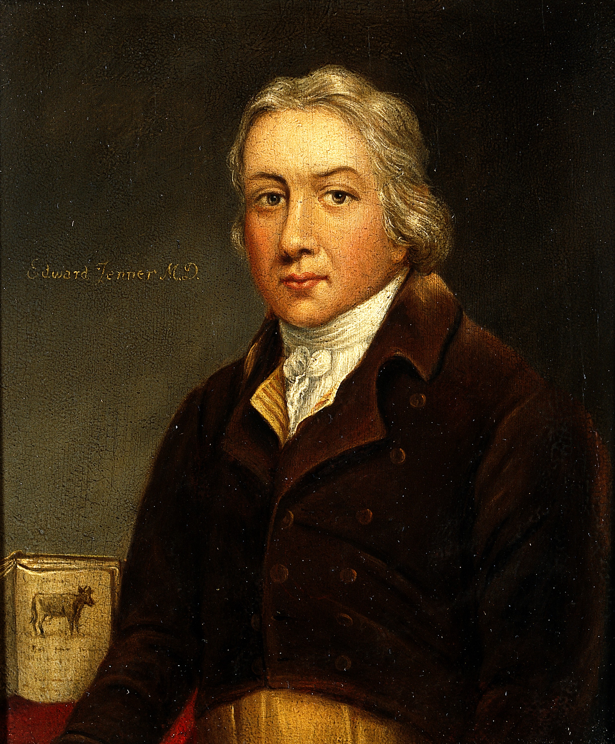
In 1768 John Fewster an English physician made a key observation. He noted that people who had been infected with cow pox appeared to having an immunity to small pox. Many replicated this, famously Benjamin Jesty used cow pox to immunise his family from small pox in the early 1770s. Jesty wasn’t a doctor so didn’t end up publishing his findings, meaning we would have to wait another 20 years before Jenner conducted his work.

Jenner put together a study taking the pus from a milkmaid, called Sarah Nelmes who had caught cowpox from a cow named Blossom. Blossom’s hide hangs in the library of St George’s medical school in London today. The pus was administered into both arms of 8 year old James Phipps. The boy experienced a mild fever and symptoms of cowpox, but recovered unharmed. Jenner then injected Phipps with variolous material for small pox. He showed no sign of infection. Follow up injections also yielded no signs of small pox. This confirmed to the scientific community that the boy had developed an immunity. This was a landmark in medical science as Jenner had proven that the method of vaccination (named from vacca meaning cow) could provide people immunity from small pox. A commercial vaccine was generated in 1798 and since its wide spread use small pox has been completely eliminated from the human population.

Edward Jenner - Wikipedia
Edward Jenner

With advances in medical technology, along with out understanding of microbiology scientists were able to develop a whole host of vaccines for problematic pathogens. Including rabies, influenza, measles, rubella, polio, tetanus the list goes on and on.

Vaccine Theory

We know that the aim of a vaccine is to generate immunity in the recipient to a specific viral or bacterial infection. Simply put vaccination follows the idea of “know your enemy” training your immune system in case it encounters the infection. I have discussed the immune system in previous posts, but I will do a small recap. We divide the immune system into two parts, which we call the innate and adaptive responses. Our innate immune system will do its best a front line defence force. Identifying foreign pathogens and eradicating them as quickly as possible. Sometimes that is not enough, some viruses and bacteria replicate too quickly or have ways to avoid detection long enough for them to cause damage. This is where the adaptive immune system comes in. These specialised cells change their tactics depending on what exactly they are fighting against.

Our adaptive immunity is constantly learning and it is this ability to learn that is utilised when administering a vaccine. The cells of the adaptive system produce structures called antibodies. These are Y shaped proteins that can be customised to suit their purpose in targeting one specific virus or bacteria. The antibodies once produced can neutralise foreign pathogens in a number of ways. They can mark them out for other cells to target and kill, disable their ability to invade other cells, or cause their death directly. However it takes time for the adaptive immune system to obtain the required information to produce these antibodies. In most cases the virus or bacteria needs to be caught and digested for valid weaknesses to be identified. We call these targets antigens, and antibodies bind to antigens. Some viruses or bacteria are able to avoid being broken down and suitable antigens targeted. This means that the adaptive immune system cannot work effectively and the infection is free to spread through the body. Viruses such as the one for HIV chooses to target immune cells directly killing them before they can kill it. Problems like evasive pathogens and the delay between infection and the production of antibodies can lead to a huge escalation in the infection leaving the body in a state it can fight back from.

The Y shaped antibody is produced by the adaptive immune system to combat foreign pathogens. The variable ends marked denoted in blue are changed to cater to different pathogen antigens.

One of the most crucial cells of the adaptive immune system are the memory cells. These as their name suggests will remember previous attacks and the types of antibody used then immediately produce the required antibodies in response. This allows the body to quickly react to an infection it has experienced before without the need to produce new antibodies.

The concept of vaccination is to therefore introduce the immune system to a pathogen in a safe manner so that it can prepare antibodies in case it encounters the real live version in the future. The way we introduce the immune system to these viruses or bacteria is through one of 4 methods.

  1. Live Attenuated: this is a weakened version of the pathogen it is unable to cause infection in healthy individuals, but will cause a very rapid immune response.
  2. Killed Whole Organism: a dead version of the pathogen.
  3. Purified Proteins or Polysaccharides: the antigens of the pathogen extracted and purified no injection of the original pathogen.
  4. Genetically Engineered: we can use genetic techniques to make the antigen without the need for the original pathogen. These can be injected safely triggering a smaller immune response.

By studying individual viruses or bacteria we can identify suitable antigen targets than can be used in vaccines for future immunisation within the human population. Vaccination has prevented innumerate deaths from diseases we no longer have to worry about. I will caution this with the worrying movement of parents not vaccinating their children. The United States last year saw large outbreaks of measles with the disease making a resurgence due to a failure to vaccinate. Through fear and misinformation people are being put at risk. The work being done today to develop a vaccine for COVID-19,which will hopefully prevent the lock-down many of us are under now from occurring again. We are living through the impact, an uncontrolled virus can have on our species. It is vaccination that has prevented that from happening with many other potentially worse pathogens. It is important to note that a vaccine takes years to fully develop with the pharmaceutics trials required before it is safe for usage with the general population. Vaccines have proven themselves to save lives time and time again. Understanding their benefit is crucial to our future.

Reading Sources

A Real Headache

Headaches are an interesting area of pain. The brain itself does not possess any receptors for detecting pain. The sensations we feel are due to changes in the muscles, blood flow and other nerves within the head. Your brain is not in pain even if it may feel that way. In fact is the areas surrounding the brain, the scalp, blood vessels and the tissue covering the brain called the meninges are sensitive to pain.

The occurrence of headaches is relatively common they are perhaps the biggest reason people seek over the counter pain medication. They have been found to affect around 15% of adults within the USA and are more common in women than men. This is due to the effect that the female hormone oestrogen has on the chemical balance within the brain.

Headaches themselves can be classified into two main types primary headaches and secondary headaches. The primary class are the ones we are perhaps most familiar with they include migraine, tension headaches and cluster headaches. The secondary are far more serious, they are usually linked to some other issue within the body hence they are a symptom of the other problem. Conditions such as cancer, or an ongoing infection that produce headaches as a symptom need to be treated immediately. Within in this post I will only be covering the primary class of headache.

Types of Primary Headache

As mentioned above there are 3 main types of primary headache as characterised by the international classification of headache disorders (there is a forth category that includes a subset of different headache types which don’t fit into these main three). They include the type 1 migraine, type 2 tension and type 3 cluster. They differ in the symptoms and the regions of the head they affect. I will cover each of these types of headache, their causes and treatments we have for them.

Migraine

There are an estimated 1 in 7 people who suffer with migraines. There are several factors that cause migraine from genetics, sex and neurological changes. Firstly it is important to note that migraines can be hereditary, passing from one generation to the next. Studies done in twins have shown a higher occurrence of migraines in patients with a family history of the disease. Next there is your sex, women are 3 times more likely to suffer with migraines than men. This is due to the hormone oestrogen. Prior to puberty in children migraines occur in similar levels in both males and females. Once puberty begins there is a higher level of oestrogen in females which leads to a higher prevalence of migraines. This has been confirmed in rat models of migraine with variations observed throughout the menstrual cycle.

So what is behind migraines? This is where the science becomes a little complicated. There are many prevailing theories over the exact cause. There exist two main camps competing with each other. The vascular approach which believes that the blood vessels surrounding the brain are responsible, and the neurological approach that say the neuronal mechanisms within the brain itself are responsible. Some are so fixated on their view that papers named “The vascular theory of migraine – a great story wrecked by the facts” have been published. However this has lead many to look at both and that it is likely migraines are caused by dysfunction in both these systems.

What we do know is that a common symptom of migraine is aura. This is the sensory disturbance patients will experience prior to and/or during a migraine headache. Changes in vision are the most common with people losing their full field of view or flicking within the images they see. This is theorised to be caused a phenomena called cortical spreading depression (CSD). CSD is a burst of nerve activity within the brain followed by a period of very low activity. This spreads like a wave across one side of the brain.

The most prominent feature of migraine is the pain. The cause of which is as to be expected still up for debate. However as I have stated the brain feels not pain, so it has to be the vessels and tissues surrounding the brain that are triggering the sensation. There are multiple areas of the brain that are involved in the pain pathways and the control of blood pressure and flow. These areas have connections between each other and it is the activation of these links that is believed to cause the pain around the head during a migraine headache attack.

The way we go about treating migraine is with a group of drugs called the triptans. The most common of which is sumatriptan. These work to constrict the blood vessels around the brain helping to reduce the pain associated with migraine. There are nasal spray versions that allow for immediate relief during a migraine attack as well as an oral tablet for prevention.

Overall a migraine is an inherited neurovascular disease that is caused by a dysfunction within the brain. Leading to irregular activation of the sensory, vascular and pain pathways causing headache.

Tension

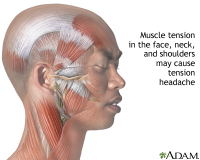
These are perhaps the most common among these three types. They are often the reason people seek pain medication at pharmacies. Unlike migraine the tension headache is muscular in origin. Usually affecting both sides of the head and generating a feeling of tightness rather than the pulsing throb of a migraine. Tension headache sufferers do not tend to show signs of light or sound sensitivity but it can occur. Nor do they show nausea or vomiting.

Tension headaches themselves are usually a catch all class, for things that don’t come under the other two classes. The best treatment for them are your common anti-inflammatory drugs such as paracetamol, ibuprofen and aspirin. These work to reduce the activity of the immune system leading to a reduction in the pain around the head.

Tension-type headache
This image shows the number of muscles that surround the head and face. When these dysfunction or receive incorrect signalling they can produce tension pain.

Cluster

The simple fact that in most cases you will see the word excruciating used to describe cluster heads, shows the level of pain they cause people. Some female sufferers have described the feeling as worse than childbirth. The cluster headache is the third and final class I will discuss. They are in a similar way to migraine neuronal in origin. They affect one side of the face causing trademark symptoms of loss of eye control (drooping of the eyelid, tear production and contraction of the pupil) and a runny nose (nasal discharge).

Specifically the trigeminal nerve. This nerve is a member of the twelve cranial nerves that all have very specific functions in the control of your senses. The trigeminal nerve connects to your face and is responsible for controlling facial movement and the sensation of touch. It branches into three distinct pathways that basically divide your face into thirds.

It is the opthalmic branch that is responsible for the pain associated with cluster headaches. All these headaches are due to a dysfunction in the way your nerves are acting or processing information. Cluster headaches generate a feeling of restlessness. Patients with migraines will usually remain still, but cluster patients will attempt any way to distract themselves from the pain. People have observed patients pacing, rocking on the spot and there are even some descriptions of people banging their own heads. As stated these attacks can last up to 3 hours and sufferers can experience multiple attacks a day making it extremely debilitating to their daily lives. The generate pattern to a cluster headache is weeks to months of attacks followed by a slow remission from months to years. The duration of these experiences has been recorded to last up to 15 years.

Image result for artist interpretation cluster headache
This is an artist impression of the feeling generated by a cluster headache. The feeling that something is metaphorically stabbing you in the eye.

The treatments for cluster heads follow in the same vein as migraines however there is also inhalation of 100% oxygen has been successful during cluster attacks. Other treatments include more blood vessel dilators like ergotamine.

Summary

Headaches are a complicated area of pain science. They involve multiple areas within the brain all communicating with each other to generate these pain states. The understanding of the underlying cause is still being investigated, but the progress that has been made of the past decade is huge. I hope this article sheds a little light on the inner workings of your head and how we work to treat it when things go wrong.

Reading Sources

Wuhan Virus Outbreak 2020 – Coronaviruses

The Wuhan virus has gripped the media for the past month since its outbreak in December 2019. Officials in China announced that 27 residents, mainly market stall owners of Huan seafood market had become sick with an unknown virus. Over the next month the virus has been spreading throughout Wuhan and neighbouring countries. With recent cases in both the USA and France.

Timeline of the Spread of the Wuhan Virus

  • Dec 31st 2019: Wuhan officials announce the outbreak within Wuhan, China. 27 residents of Wuhan with 7 in critical condition.
  • Jan 3rd 2020: There are now 44 confirmed patients and another 5 in Hong Kong. Scientists rule out that this is influenza or avian flu.
  • Jan 5th 2020: 59 confirmed cases and now 21 in Hong Kong. The possibility of the resurgence of SARS is dismissed.
  • Jan 9th 2020: The world health organisation (WHO) confirm a novel coronavirus isolated from a hospitalised patient.
  • Jan 10th 2020: The virus claims its first victim; a man in Wuhan.
  • Jan 13th 2020: First case in Thailand.
  • Jan 16th 2020: Chinese national hospitalised in Japan confirmed to be the same virus after the individual was likely in contact with an affect person.
  • Jan 21st-23rd 2020: First cases in the USA in Washington and Chicago.
  • Jan 22nd 2020: China suspends all travel from Wuhan placing the city under quarantine.
  • Jan 24th 2020: First confirmed case in France.
  • Jan 26th 2020: Two more cases of the virus in California and one in Arizona.
  • Jan 30th 2020: First confirmed case in Tibet. With cases now confirmed in all 21 provinces of mainland China.

Introduction to Viruses

The reason I want to talk about this outbreak is to explain how these viruses work and what is the best way for us to help prevent its spread. Viruses are unique, they are in the most simplistic way a living organism. Although they possess no cellular structure or need to gain sustenance. They do contain genetic material that allows them to reproduce and evolve.

Viruses are defined in many ways (for more information search for the Baltimore Classification), but here I will mention only DNA and RNA containing viruses. In the situations I will discuss their functions are identical. DNA and RNA are both genetic instructions, that hold information for the creation of more virus.

Viruses are unable to self-replicate or undergo sexual reproduction. They instead require a host cell to conduct this for them. Once a virus enters the body it will infect the hosts cells injecting within them the DNA or RNA that they are carrying. The internal machinery of our cells will read this foreign genetic material and use it to make more of the virus. Our cells are relatively simple in this regard. Provide them information and they will make it no questions asked. So our cells unwittingly go about their normal function leading to the production of more viruses. This eventually leads the cell to die allowing the new born viruses to escape to infect further cells down the line.

Treating Viruses

This life-cycle makes viruses a tricky area to treat medically. The common cold has no cure and treatments for HIV have taken years to be developed. The reason for this is that the virus spends a large amount of its time within the host’s cells. This effectively hides them from the drugs that could target them. We struggle to make drugs that are able to differentiate between what is normal and infected. The same obstacle cancer treatments have. Therefore in many situations involving viruses we rely on the immune system to handle the problem. Conditions such as chicken pox or colds are are left up to the individual to suffer through. Only using methods to stem the symptoms such as creams for chicken pox and anti-inflammatory drugs for colds.

That’s not to say there aren’t antiviral drugs. These target the life cycle of the virus in question working to prevent their reproduction. They are however specifically designed for the virus in question with treatments existing for HIV and hepatitis.

Coronavirus

That brings us to the topic of the month coronaviruses. This is actually the name of a family of viruses. From the common cold to influenza to SARS (server acute respiratory syndrome) and the recent novel coronavirus 2019 (nCoV) of Wuhan. It is important to note that there is not much known of this new virus. Therefore things are subject to change, any numbers or statistics posted are accurate from today, but will change in the coming months.

The origin of the virus is believed to have been a jump from animal to human with the suspects being either bats or snakes. It may have been contracted through being in close proximity to these animals carrying the virus or consumption of its meat. There are several factors to take into account when looking at a new virus. These are its method of transmission and the incubation period. With regards to the nCoV it is air-born infecting others through bodily fluids, coughing and sneezing. Inhaling the infected air will allow the virus into the lungs where it will begin replicating. The incubation period is the time it takes for symptoms of the virus to become noticed. Once the virus is in your system it will not immediately cause fever and coughing only once it had spread enough throughout the lungs will these begin to show. For the nCoV this period is currently estimated at 7-14 days, this is the reason why those escaping Wuhan and mainland China are being kept in quarantine for a minimum of two weeks.

Thus far we know the virus causes similar symptoms to that of flu; fever, coughing and in extreme circumstances pneumonia. There is currently no cure to the nCoV with hospitals only able to offer supportive care to those in need. Providing medication for the symptoms, fluids to those undergoing a fever and in some cases respiratory support for patients struggling to breathe. There is now ongoing work to develop a vaccine for the nCoV in the USA. The national institute of health announced their work on January 28th.

It is important not to panic, the WHO have not yet classed this outbreak as a global pandemic. This means it is crucial that people remain vigilant and alert without becoming overly anxious about this subject. This outbreak is not a crisis that requires mass worry. If you are concerned, the WHO and the centres for disease control and prevention (CDC) have provided a list of recommended actions to prevent the transmission and spread of this virus.

  1. Wash your hands often with soap and water for a minimum of 20 secs. Or using alcohol based sanitizer with a least 60% alcohol.
  2. Avoid touching your face with unwashed hands when outside. Viruses can enter the body through the nose, mouth and eyes.
  3. Avoiding contact with those who are already sick. As well as remaining home when you yourself are sick.
  4. Make sure to cover your mouth with a tissue should you cough or sneeze and dispose of the tissue into the rubbish. With regard to masks; they will help to prevent people who are already sick spreading their virus too easily. However their effectiveness at preventing the inhalation of infected air is relatively minimal.
  5. Avoid unnecessary travel to infected countries. Placing yourself in a higher risk area will increase chances of contracting the virus.
  6. If you are concerned you may have the nCoV contact your local medical professional over the phone. They will then be able to prepare correctly to deal with your condition. However it is unlikely you have the nCoV unless you have been in direct contact with someone who has recently travelled from China.

As of January 29th 2020 there have been 7815 confirmed cases of the nCoV with 170 deaths (only within mainland China). Compared to the SARS outbreak of 2002-03 with 8098 cases and 774 deaths, the nCoV does not appear to have the same lethality however it is still within all recommendations to avoid its spread any further.

Reading Sources

Addiction – A New Years Resolution Staple

A new year, a new you. As December passes into January and we once again have to remember to put 2020 instead of 2019 on paperwork. We often decide to make a change in our lives, one for the better. One of these more common resolutions is to quit smoking. Now I don’t need to tell you that inhaling high temperature toxic gas causes damage, but instead talk about how the brain becomes addicted to these bad habits.

Sorry I lied, I can’t not give a general warning. Smoking causes cancer, damages the lungs and trachea as well as leading to discolouration of the skin and rapid ageing. Not a good habit best we get rid of it. Vaping won’t be left to its own devices either. Dosages of nicotine alone is also no better for you.

This post will aim to focus on addiction. Using nicotine as the example, this chemical is the reason people becoming hooked on smoking and more recently vaping. We will delve into how the brain is able to develop a dependence on toxic chemicals.

Nicotine

The addictive component of conventional and electronic cigarettes. Nicotine is able to enter the brain and cause all manner of issues throughout your system. There are many natural chemical signals within the brain that all conduct specific functions; from the control of your emotions, the processing of pain and the integration of your muscles. One of these chemical signals is the neurotransmitter acetylcholine. Nicotine happens to be able to conduct the same function as acetylcholine activating different areas of the brain outside of your control. In fact nicotine is so good at this that scientists named a group of acetylcholine receptors (detectors of acetylcholine) as nicotinic. Nicotine is able to turn on these receptors without acetylcholine being present.

Nicotine has some minor benefits in the form of improved short term memory and increased focus. However these benefits do not outweigh the increased chances of cancer, increased blood pressure, along with other cardiovascular complications, and eventually dependence.

Reward

The reward pathway is found within the brain and acts to promote actions that improve our health and keep us going. Things such as eating, sex and social interactions all work toward your basic instincts to survive and procreate. Whenever you engage in these activities the brain rewards you with feelings of pleasure. The areas of the brain responsible of this are, the ventral tegmental area (VTA) and the nucleus accumbens (NA), together they make up this “reward” pathway. When activated they will release dopamine which generates the feelings of euphoria. This in turn promotes you to repeat those actions reinforcing it into your memory.

A simplified diagram of the brain regions involved in the reward pathway. Stimulation of the ventral tegmental area sends signals causing the release of dopamine into the frontal cortex. This generates the feeling of pleasure associated with reward.

Mechanisms that Drive Dependence

Abusive drugs take advantage of this system tapping into the potential of reward to develop addiction. Along with all the negatives; they provide that one positive people crave, pleasure! Nicotine being our example of the day, is able to activate the reward pathway by binding to acetylcholine receptors within the VTA. Carrying out the function that internal acetylcholine would have conducted. This leads to the NA releasing dopamine effectively rewarding you for something that is causing you harm. This is what leads to the development of addiction.

By repeatedly engaging in the drug activity, such as smoking the inhaled nicotine continues to promote dopamine release. The brain eventually becomes accustomed to the constantly higher level of dopamine, therefore more of the drug is required to achieve the same levels of pleasure. Through the usage of the drug the addict has reprogrammed their brain into thinking these high levels of dopamine are normal. This means that the act of quitting is even more difficult.

Quitting Addiction

As simple as it may seem to an outsider, quitting is not an easy undertaking. To remove the new programming your brain has developed over the usage of the drug is extremely taxing. There is now an innate imbalance, in the chemicals within the brain. It is therefore advised in most cases to use medicinal help, to slowly reduce cravings and help with withdrawal. Going “cold turkey” can cause all manner of issues both mentally and physically, some may have experienced these effects when failing to get their morning coffee. Headaches and irritability are just a few of the potential withdrawal symptoms one might experience when quitting an addiction. Medicinal help for nicotine dependence consists of using nicotine patches or alternative nicotine replacement. Then to eventually reduce the dose over time, allowing you to correct the chemical imbalance without too much of the craving or withdrawal. This takes time, the brain has completely changed how it responds acts of pleasure. It needs to slowly repair the damage the drug has caused to its internal circuits. The best method to quit any drug is to seek medical consultation and to develop a regime works best for you.

Summary

Good luck to those who are trying to make a positive change in their life, I shall leave you with a quote and a clip from Dr Robert Lustig. These drugs and substances provide pleasure not contentment or happiness in your life. “Pleasure is short lived and happiness is long lived.” Seek happiness for a better state of mind and a Happy New Year!

Reading Sources

  • Molecular and Cellular Aspects of Nicotine Abuse, John Dani and Steve Heinemann.
  • Drug addiction: the yin and yang of hedonic homeostasis, Koob GF.
  • Neurobiological mechanisms of addiction: Focus on corticotropin releasing factor, George Koob and Eric Zorrilla.
  • Effect of nicotine on brain activation during performance of a working memory task, Ernst et al.
  • Reward, Addiction, Withdrawal to Nicotine, Mariella de Biasi and John A. Dani.

A Return to Replacement

This will be the follow up to my previous post on the history of transplantation. Here I will cover the current method of transplantation along with the negatives that come with it. Then I will move to discussing the future of transplantation along with methods that could make traditional methods obsolete.

Current Methods

Transplants are pretty commonplace these days, through the surgical advances made throughout the 20th and 21st centuries. Such things as better equipment, higher sterility during surgery and the development of drugs to prevent rejection. There are still several caveats to transplant surgery that make it almost an art in finding that perfect match.

There are two primary requirements than need to be met for a donor to be considered. The blood types much match or there will be immediate rejection the positive or negative nature of the blood does not matter however.

As shown here, people with blood type O are universal donors, and people with AB blood are universal recipients.

Secondly the more complicated requirement is a need for matching human leukocyte antigens (HLA). These are special proteins present on all cells in your body that determine what is you compared to what is foreign. If these do not match your immune system will immediately recognise the transplanted tissue as foreign and act to destroy it. Also in some conditions, as mentioned in my previous post, the possibility of the transplant attacking the donor. It is not feasible to acquire a perfect match unless dealing with twins however doctors try to obtain as close a match as is available. This is far more complex than blood matching as there are many different HLA markers on a singular cell to consider. In most cases doctors look to match 8-10 of these markers to a potential donor. Finding a match is far more likely when looking within family as markers are past on from generation to generation. This does not rule out outside donors of course.

Along with finding the necessary match we come across several other issues. The availability of organs is limited and some organs can only be obtained from a deceased donor e.g. the heart. For an organ like the kidney the waiting period can be years and with 6000+ people in the UK alone (NHS Statistics 2019) on the waiting list for transplant, time is limited.

If you do manage to go through surgery, you will need to be put on immuno-suppressant drugs to prevent your immune system from attacking your newly transplanted organ. This is a lifetime commitment which obviously comes with its own risks. By constantly suppressing your immune system you become more susceptible to disease and illness. Now I am not saying that transplantation is a bad thing. It is an incredible leap in our medical history and many, many lives have been saved. Today I am going to talk about how science is aiming to improve this technique. In many cases moving away from the need to receive organs from others and removing the need for donated tissue at all.

Growing Your Own

You would think it science fiction, but we have come a long way. Here I will discuss methods being developed to potentially grow organs for transplantation as well as making our own replacement tissues. This will lightly cover not only transplantation, but the ways we can repair systems without the need for full organ transplants. I will not be covering prosthesis used in amputations, that is an area all to itself.

The key to much of the advances in replacement technology and research are stem cells. You will hear mention of stem cells through this post so a quick, simple explanation will be needed. Stem cells are unique cells with their main property being the ability to become any cell in the body given the right stimulus. They are basically blank sheets with possibility to become anything for example, heart cells, muscle cells or liver cells. These cells hold within them the potential to become entire organs if treated correctly. They are found throughout your body and can be extracted and purified by labs and it is here where our journey to lab grown organs begin.

Future of Skin Grafts

Skin grafts included one of the first historically documented forms of transplant (circa 16th Century). They are a crucial part of many surgeries and in the treatment of several conditions such as burns. For burn victims the treatment is often times just as bad if not worse than receiving the burn in the first place. Many treatment options involve the use of allografts. Skin taken from one area of the body and used to cover another, this will lead to the formation of two wounds that need to be cared for. So studies have been done using stem cells to treat wounds. These stem cells can become new skin cells to eventually help repair that which has been damaged. These applications have been shown to accelerate the wound healing process. Not only repairing the lost skin, but restoring the hair follicles, sebaceous glands and overall improving the look of the area. This improves the psychological state of the patient along with the advanced healing. The treatment has shown such success that some groups have developed stem cell guns that can spray the cells onto the burnt regions.

It’s not just burns that can be treated with steam cells. In 2017 an article was published in Nature documenting the treatment of a young patient with junctional epidermolysis bullosa (JEB). This is a genetic condition causing skin to blister and become damaged easily. Generically modified steam cells grown in the lab were applied to the skin of the patient. These over time eventually replaced the entire epidermis.

Let’s take this further, how about growing substitute skin. One company has developed a material called Stratagraft, a skin substitute entirely grown from living cells in the lab that can be used successfully in transplants. Stratagraft is made from living cells that scientists aim to be universal. To avoid the problem of rejection the tissue is made from keratinocytes. These are the most prevalent cell found in the skin and do not express one of the main markers for the immune system to recognise as foreign. Along with this precaution the fabricated tissue contains no immune cells themselves. The tissue when grown forms a layer almost identical to that of normal human skin. Stratagraft has been used in clinical trials, and is showing great promise in the treatment of injuries that would require skin transplants.

Cellular Construction

The dream of growing organs is a little way off, but there are many who are attempting new and unique approaches to achieving this goal. When we grow cells in the lab they tend to form simple flat structures. Cultured cells will not form three dimensional organs and on top of that organs are not simply made from one cell type. There are a variety of different cells that make up the heart along with the need to form chambers and vessels. These things are complicated and sure we can grow the individual cells that make up a heart but we can’t tell them to make one.

Some groups have looked to scaffolds, creating a skeleton for the cells to grow on and form organs. Again organs are not simple so require so much work to even get close to a functional tissue. However there has been some success in using scaffolds to grow retinal transplants. The technology is very young and the work is still aiming to find the perfect material for implantation. Testing whether is it better to use a degradable scaffold that will eventually disappear or a permanent one. In fact a retinal patch was due to enter phase 3 clinical trials this year after early success in two patients suffering from macular degeneration (blindness due to the loss of a layer in the eye that provides nutrients within the retina). Some say that the use of a scaffold over complicates the surgery, but they are going forward with their patch. Some day treating blindness due to age might be as simple as getting a new retinal layer transplanted.

I cannot really avoid talking about scaffolds without mentioning the controversial surgeries that took place back in 2011. The world was in awe at the miraculous treatment performed by Dr. Paolo Macchiarini. The first ever tracheal transplant successfully completed using an entirely synthetic windpipe. However it was not joy for too long. Andemariam Beyene lived for 3 years after the surgery and was the longest surviving patient of this surgery. Several other transplants were performed all leading to the deaths of the patients. Those who survived had their synthetic windpipes removed in replacement of a living donor version. Macchiarini seemed ignorant of these failures and continued his work in clinical trials in Russia. Investigations were underway unearthing false research claims in past publications and medical negligence on the part of Macchiarini. Sadly no crime could be proven as the patients may have died under any other treatment. The Swedish national scientific review board did find misconduct in his publications and had his papers retracted. So why didn’t these synthetic windpipes work? They couldn’t integrate correctly with the body, causing inflammation of the living windpipe, and although they were sown with living cells no true tissue really grew back.

Making Miniature Organs

Let us move to the really exciting advances. We have covered replacement methods and using scaffolds with stem cells. So it’s time to talk about making organs and tissues ourselves. Dr Jim Wells and his team have put together methods of taking stem cells and coaxing them to become specific cells and then work together to form effectively miniature organs called organoids. These are very small and around the size of a pea, but they stand as a huge step in developing methods that could eventually grow organs. Currently they are incredibly useful to medical research and patient treatment in rather unique ways. These organoids possess much of the complexity of the original organ, they possess various cell types and are layered. They also have the same functions as the organ they are derived from.

This functionality makes them ideal for the testing of drugs. Many drugs fail to reach the level of clinical trial or fail before getting to consumers approximately 90% of drugs in fact fail. Drug development takes many years of research and much of the early testing goes on in animals. However we are humans not rats or monkeys we are all genetically different. A drug that may treat arthritis in a rat may have no effect on a human. So these organoids stand as a method of reducing animal testing as well as having an artificial human test subject for new therapeutic treatments. Not only that but these organoids have been used to create personalised treatments for patients. Taking a sample of the patient’s stem cells allows the growth of an organoid unique to them. Drugs can then be tested without any harm to the patient, generating individual treatment plans.

Bioprinting

With the rise of 3D printing within the manufacturing industry. Along with the ability for people to craft things from desktop printers, this technology has leapt forward in the past few years. It is now the turn of scientists to jump on the bandwagon. Swapping out the plastics for bio-ink. There is now a huge amount of investment into the development of bioprinters that have the capability of printing cells to form shapes and 3D structures similar to blood vessels and the trachea. The cells are threaded together within a string the size of a hair. The string is then layered to form these structure. They have produced replicas of an asthmatic trachea that will relax and contract in response to irritation and drug therapy. This technology along with the organoids can also be used highly within the drug development field. In a few years time we may be able to print new tissue to replace damaged organs or at least parts of organs together with blood vessels.

Summary

The area of transplantation is ever changing we started back in the 1900s with the first successful kidney transplant and now we perform surgery with ease. The next hurdles for us to cross are the problems of rejection and the limited supply of organs for those in need. The advancement of this technology aims to remove rejection as a problem by providing the patient’s own cells as transplants and then help with the supply by growing replacement organs in the future. Some groups are even looking into using donor organs and removing all the cells from it leaving behind only the nature scaffold then filling it with the recipient’s cells, as a sort of stop gap between these methods I have discussed. We also have advancements in the way we are conducting drug development using human based tissues as a replacement for animals. In a couple more years who knows how things may have advanced. The future of bio-engineering is truly exciting and one day we may be waiting only a month to grow a new kidney that is our very own.

Reading Sources

  • The future of organ transplantation. Cascalho. M et al.
  • Individual cell-only bioink and photocurable supporting medium for 3D printing and generation of engineered tissues with complex geometries.
  • Bioengineered stem cells as an alternative for islet cell transplantation. Moore S. et al.
  • Scaffolds for retinal pigment epithelial cell transplantation in age-related macular degeneration. White and Olabisi.
  • Evaluation of culture conditions for in vitro meniscus repair model systems using bone marrow-derived mesenchymal stem cells. Hidalgo Perea, et al.
  • Scaffold-free trachea regeneration by tissue engineering with bio-3D printing. Taniguchi et al.
  • Tissue-engineered trachea from a 3D-printed scaffold enhances whole-segment tracheal repair. Gao et al.
  • Bioartificial Organ Manufacturing Technologies. Xiaohong Wang.
  • Stem Cells for Skin Tissue Engineering and Wound Healing. Chen et al.
  • Regeneration of the entire human epidermis using transgenic stem cells. Hirsch et al.
  • Stratagraft clinical trials
  • StrataGraft skin substitute is well-tolerated and is not acutely immunogenic in patients with traumatic wounds: results from a prospective, randomized, controlled dose escalation trial. Centanni et al.
  • The effects of stem cells on burn wounds: a review. Francis et al.
  • Stem Cell Treatment in Retinal Diseases: Recent Developments. Ayşe Öner.